
Page 103 - Atlas of Small Animal CT and MRI
P. 103
Globe 93
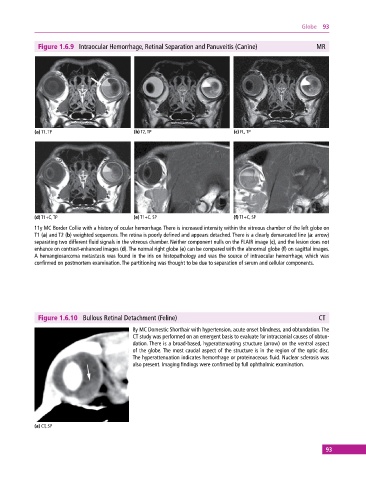
Figure 1.6.9 Intraocular Hemorrhage, Retinal Separation and Panuveitis (Canine) MR
(a) T1, TP (b) T2, TP (c) FL, TP
(d) T1+C, TP (e) T1+C, SP (f) T1+C, SP
11y MC Border Collie with a history of ocular hemorrhage. There is increased intensity within the vitreous chamber of the left globe on
T1 (a) and T2 (b) weighted sequences. The retina is poorly defined and appears detached. There is a clearly demarcated line (a: arrow)
separating two different fluid signals in the vitreous chamber. Neither component nulls on the FLAIR image (c), and the lesion does not
enhance on contrast‐enhanced images (d). The normal right globe (e) can be compared with the abnormal globe (f) on sagittal images.
A hemangiosarcoma metastasis was found in the iris on histopathology and was the source of intraocular hemorrhage, which was
confirmed on postmortem examination. The partitioning was thought to be due to separation of serum and cellular components.
Figure 1.6.10 Bullous Retinal Detachment (Feline) CT
8y MC Domestic Shorthair with hypertension, acute onset blindness, and obtundation. The
CT study was performed on an emergent basis to evaluate for intracranial causes of obtun-
dation. There is a broad‐based, hyperattenuating structure (arrow) on the ventral aspect
of the globe. The most caudal aspect of the structure is in the region of the optic disc.
The hyperattenuation indicates hemorrhage or proteinaceous fluid. Nuclear sclerosis was
also present. Imaging findings were confirmed by full ophthalmic examination.
(a) CT, SP
93