
Page 104 - Atlas of Small Animal CT and MRI
P. 104
94 Atlas of Small Animal CT and MRI
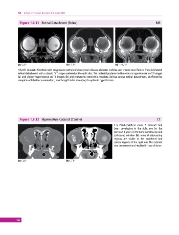
Figure 1.6.11 Retinal Detachment (Feline) MR
(a) T2, TP (b) T1, TP (c) T1+C, TP
18y MC Domestic Shorthair with progressive central nervous system disease, diabetes mellitus, and chronic renal failure. There is bilateral
retinal detachment with a classic “V” shape centered at the optic disc. The material posterior to the retina is hyperintense on T2 images
(a) and slightly hyperintense on T1 images (b) and represents retroretinal exudate. Bullous serous retinal detachment, confirmed by
complete ophthalmic examination, was thought to be secondary to systemic hypertension.
Figure 1.6.12 Hypermature Cataract (Canine) CT
11y Poodle/Maltese cross. A cataract had
been developing in the right eye for the
previous 4 years. In the bone window (a) and
soft‐tissue window (b), mineral attenuating
regions are visible in the peripheral and
central regions of the right lens. The cataract
was brunescent and resulted in loss of vision.
(a) CT, TP (b) CT, TP
94