
Page 102 - Atlas of Small Animal CT and MRI
P. 102
92 Atlas of Small Animal CT and MRI
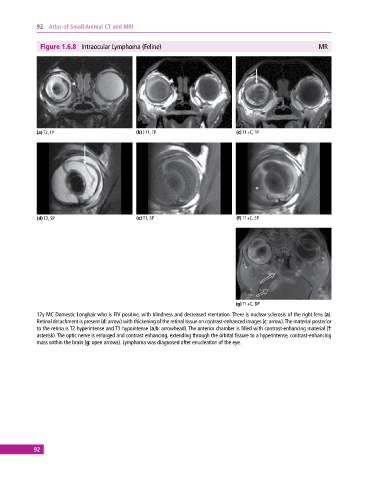
Figure 1.6.8 Intraocular Lymphoma (Feline) MR
(a) T2, TP (b) ) T1, TP (c) T1+C, TP
(d) T2, SP (e) T1, SP (f) T1+C, SP
(g) T1+C, DP
12y MC Domestic Longhair who is FIV positive, with blindness and decreased mentation. There is nuclear sclerosis of the right lens (a).
Retinal detachment is present (d: arrow) with thickening of the retinal tissue on contrast‐enhanced images (c: arrow). The material posterior
to the retina is T2 hyperintense and T1 hypointense (a,b: arrowhead). The anterior chamber is filled with contrast‐enhancing material (f:
asterisk). The optic nerve is enlarged and contrast enhancing, extending through the orbital fissure to a hyperintense, contrast‐ enhancing
mass within the brain (g: open arrows). Lymphoma was diagnosed after enucleation of the eye.
92