
Page 105 - Atlas of Small Animal CT and MRI
P. 105
Globe 95
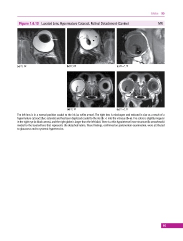
Figure 1.6.13 Luxated Lens, Hypermature Cataract, Retinal Detachment (Canine) MR
(a) T2, DP (b) T2, DP (c) T1+C, TP
(d) T2, TP (e) T1+C, TP
The left lens is in a normal position caudal to the iris (a: white arrow). The right lens is misshapen and reduced in size as a result of a
hypermature cataract (b,c: asterisk) and has been displaced caudal to the iris (b: >) into the vitreous (b–e). The sclera is slightly irregular
in the right eye (e: black arrow), and the right globe is larger than the left (d,e). There is a thin hypointense linear structure (b: arrowheads)
medial to the luxated lens that represents the detached retina. These findings, confirmed on postmortem examination, were attributed
to glaucoma and to systemic hypertension.
95