
Page 110 - Atlas of Small Animal CT and MRI
P. 110
100 Atlas of Small Animal CT and MRI
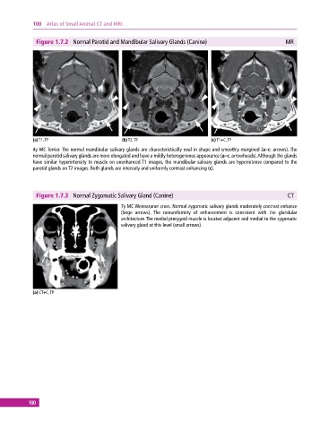
Figure 1.7.2 Normal Parotid and Mandibular Salivary Glands (Canine) MR
(a) T1, TP (b) T2, TP (c) T1+C, TP
4y MC Terrier. The normal mandibular salivary glands are characteristically oval in shape and smoothly margined (a–c: arrows). The
normal parotid salivary glands are more elongated and have a mildly heterogeneous appearance (a–c: arrowheads). Although the glands
have similar hyperintensity to muscle on unenhanced T1 images, the mandibular salivary glands are hyperintense compared to the
parotid glands on T2 images. Both glands are intensely and uniformly contrast enhancing (c).
Figure 1.7.3 Normal Zygomatic Salivary Gland (Canine) CT
7y MC Weimaraner cross. Normal zygomatic salivary glands moderately contrast enhance
(large arrows). The nonuniformity of enhancement is consistent with the glandular
architecture. The medial pterygoid muscle is located adjacent and medial to the zygomatic
salivary gland at this level (small arrows).
(a) CT+C, TP
100