
Page 115 - Atlas of Small Animal CT and MRI
P. 115
Salivary Glands 105
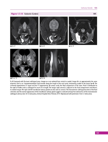
Figure 1.7.13 Sialocele (Canine) MR
(a) T2, TP (b) T2, TP (c) T2, TP
(d) T1+C, TP (e) T2, DP
5y M Samoyed with fluctuant sublingual mass. Images a–c are ordered from rostral to caudal. Image d is at approximately the same
anatomic level as a. Compartmentalized fluid extends along the length of the oral cavity, terminating caudal to the larynx (a–d). The
uniformly hyperintense T2 signal and the T1 hypointensity (d: arrow) verify the fluid composition of the mass. Fluid is distributed to
the right of midline and is sublingual for much of its length. The tongue (a,b: arrows) is adjacent to the fluid compartment and distorts
its medial margin. The right and left mandibular salivary glands are also seen (c: arrows). The characteristic sublingual location of the fluid
collection and the normal appearance of the mandibular salivary gland suggest a sialocele arising from obstruction of the monostomatic
sublingual salivary duct. Dr S.Cizinauskas, Animal Hospital Aisti, Finland, 2014. Reproduced with permission from S. Cizinauskas.
105