
Page 112 - Atlas of Small Animal CT and MRI
P. 112
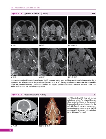
102  Atlas of Small Animal CT and MRI


              Figure 1.7.6  Zygomatic Sialadenitis (Canine)                                               MR


             (a) T1, TP                       (b) T2, TP                       (c) T1+C+FS, TP


             (d) T1, DP                       (e) T2, DP                       (f) T1+C+FS, DP
             9y FS Cocker Spaniel with left‐sided exophthalmos. The left zygomatic salivary gland (a–f: large arrow) is markedly enlarged and is T1
             hypointense and T2 hyperintense to the contralateral gland (a–c: small arrow). The contrast‐enhanced images reveal that the glandular
             architecture is retained, including the arborizing ductal pattern, suggesting diffuse inflammation rather than neoplasia. Clinical signs
             resolved with antibiotic and anti‐inflammatory therapy.


              Figure 1.7.7  Parotid Sialoadenitis (Canine)                                                 CT

                                                                              1y MC Pembroke Welsh Corgi with pain on
                                                                              palpation of left ear. The left parotid salivary
                                                                              gland, ventral and lateral to the ear canal,
                                                                              is enlarged and thickened compared to the
                                                                              right side. There is intense contrast enhance-
                                                                              ment on transverse images (a: arrows), which
                                                                                correlates with the contrast‐enhanced gland
                                                                              on the 3D images (b: arrows).


             (a) CT+C, T                      (b) CT+C, 3D, VENT

           102