
Page 114 - Atlas of Small Animal CT and MRI
P. 114
104 Atlas of Small Animal CT and MRI
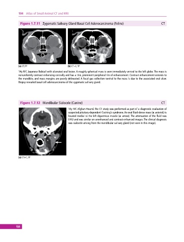
Figure 1.7.11 Zygomatic Salivary Gland Basal Cell Adenocarcinoma (Feline) CT
(a) CT, TP (b) CT+C, TP
14y MC Japanese Bobtail with ulcerated oral lesion. A roughly spherical mass is seen immediately ventral to the left globe. The mass is
nonuniformly contrast enhancing centrally and has a thin, prominent peripheral rim of enhancement. Contrast enhancement extends to
the mandible, and mass margins are poorly delineated. A focal gas collection ventral to the mass is due to the associated oral ulcer.
Biopsy revealed basal cell adenocarcinoma of the zygomatic salivary gland.
Figure 1.7.12 Mandibular Sialocele (Canine) CT
10y MC Afghan Hound. The CT study was performed as part of a diagnostic evaluation of
suspected pituitary‐dependent Cushing’s syndrome. An oval fluid‐dense mass (a: asterisk) is
located medial to the left digastricus muscle (a: arrow). The attenuation of the fluid was
0 HU and was similar on unenhanced and contrast‐enhanced images. The clinical diagnosis
was sialocele arising from the mandibular salivary gland (not seen in this image).
(a) CT+C, TP
104