
Page 119 - Atlas of Small Animal CT and MRI
P. 119
Lymph Nodes 109
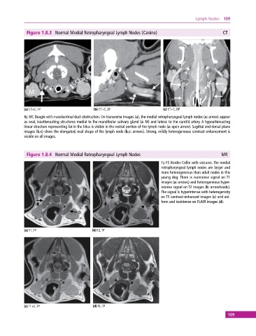
Figure 1.8.3 Normal Medial Retropharyngeal Lymph Nodes (Canine) CT
(a) CT+C, TP (b) CT+C, SP (c) CT+C, DP
8y MC Beagle with nasolacrimal duct obstruction. On transverse images (a), the medial retropharyngeal lymph nodes (a: arrow) appear
as oval, isoattenuating structures medial to the mandibular salivary gland (a: M) and lateral to the carotid artery. A hypoattenuating
linear structure representing fat in the hilus is visible in the rostral portion of the lymph node (a: open arrow). Sagittal and dorsal plane
images (b,c) show the elongated, oval shape of the lymph node (b,c: arrows). Strong, mildly heterogeneous contrast enhancement is
visible on all images.
Figure 1.8.4 Normal Medial Retropharyngeal Lymph Nodes MR
1y FS Border Collie with seizures. The medial
retropharyngeal lymph nodes are larger and
more heterogeneous than adult nodes in this
young dog. There is isointense signal on T1
images (a: arrows) and heterogeneous hyper-
intense signal on T2 images (b: arrowheads).
The signal is hyperintense with hetero geneity
on T1 contrast‐enhanced images (c) and uni-
form and isointense on FLAIR images (d).
(a) T1, TP (b) T2, TP
(c) T1+C, TP (d) FL, TP
109