Page 121 - Atlas of Small Animal CT and MRI
P. 121

Lymph Nodes  111


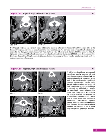
              Figure 1.8.8  Regional Lymph Node Metastasis (Canine)                                       CT


            (a) CT+C, TP                     (b) CT+C, TP
            8y MC Labrador Retriever with previously excised right tonsillar squamous‐cell carcinoma. Representative CT images are at the level of
            the medial retropharyngeal lymph nodes and are ordered from rostral to caudal. The left medial retropharyngeal lymph node (a: arrow)
            is normal in size, shape, and contrast enhancement. The central linear filling defect represents the normal fat‐filled lymph node hilus. The
            right medial retropharyngeal lymph node is markedly enlarged and has irregular margins (b: arrow). Multiple parenchymal contrast filling
            defects are characteristic of lymph node metastatic deposits. Aspiration cytology of the right medial retropharyngeal node confirmed
            metastatic squamous cell carcinoma.


              Figure 1.8.9  Regional Lymph Node Metastasis (Canine)                                       CT

                                                                             7y MC Springer Spaniel cross with previously
                                                                             excised right tonsillar squamous cell carci-
                                                                             noma. Representative unenhanced (a,b) and
                                                                             contrast‐enhanced (c,d) images are at the
                                                                             level of the medial retropharyngeal lymph
                                                                             nodes and are ordered from rostral to caudal.
                                                                             The right medial retropharyngeal lymph node
                                                                             (a–d: arrow) is markedly enlarged and irregu-
                                                                             larly shaped, has mildly indistinct margins,
                                                                             and nonuniformly contrast enhances. Filling
                                                                             defects, most clearly seen in (d), are indica-
            (a) CT, TP                       (b) CT, TP
                                                                             tive of nodal metastasis. By comparison, the
                                                                             left medial retropharyngeal node (a,c: arrow-
                                                                             head) is normal in size, shape, and contrast
                                                                             enhancement   characteristics.  Aspiration
                                                                             cytology of the right medial retropharyngeal
                                                                             node confirmed metastasis of the tonsillar
                                                                             squamous cell carcinoma as well as moder-
                                                                             ate plasmacytic and lymphocytic reactivity.


            (c) CT+C, TP                     (d) CT+C, TP


                                                                                                             111
   116   117   118   119   120   121   122   123   124   125   126