
Page 122 - Atlas of Small Animal CT and MRI
P. 122
112 Atlas of Small Animal CT and MRI
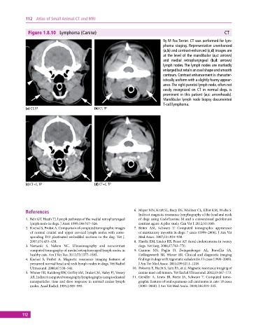
Figure 1.8.10 Lymphoma (Canine) CT
9y M Fox Terrier. CT was performed for lym-
phoma staging. Representative unenhanced
(a,b) and contrast‐enhanced (c,d) images are
at the level of the mandibular (a,c: arrows)
and medial retropharyngeal (b,d: arrows)
lymph nodes. The lymph nodes are markedly
enlarged but retain an oval shape and smooth
contours. Contrast enhancement is character-
istically uniform with a slightly foamy appear-
ance. The right parotid lymph node, often not
easily recognized on CT in normal dogs, is
prominent in this patient (a,c: arrowheads).
Mandibular lymph node biopsy documented
T‐cell lymphoma.
(a) CT, TP (b) CT, TP
(c) CT+C, TP (d) CT+C, TP
References 6. Mayer MN, Kraft SL, Bucy DS, Waldner CL, Elliot KM, Wiebe S.
Indirect magnetic resonance lymphography of the head and neck
1. Belz GT, Heath TJ. Lymph pathways of the medial retropharyngeal of dogs using Gadofluorine M and a conventional gadolinium
lymph node in dogs. J Anat. 1995;186:517–526. contrast agent: A pilot study. Can Vet J. 2012;53:1085.
2. Kneissl S, Probst A. Comparison of computed tomographic images 7. Reiter AM, Schwarz T. Computed tomographic appearance
of normal cranial and upper cervical lymph nodes with corre- of masticatory myositis in dogs: 7 cases (1999–2006). J Am Vet
sponding E12 plastinated‐embedded sections in the dog. Vet J. Med Assoc. 2007;231:924–930.
2007;174:435–438. 8. Hardie EM, Linder KE, Pease AP. Aural cholesteatoma in twenty
3. Nemanic S, Nelson NC. Ultrasonography and noncontrast dogs. Vet Surg. 2008;37:763–770.
computed tomography of medial retropharyngeal lymph nodes in 9. Cannon MS, Paglia D, Zwingenberger AL, Boroffka SA,
healthy cats. Am J Vet Res. 2012;73:1377–1385. Hollingsworth SR, Wisner ER. Clinical and diagnostic imaging
4. Kneissl S, Probst A. Magnetic resonance imaging features of findings in dogs with zygomatic sialadenitis: 11 cases (1990–2009).
presumed normal head and neck lymph nodes in dogs. Vet Radiol J Am Vet Med Assoc. 2011;239:1211–1218.
Ultrasound. 2006;47:538–541. 10. Pokorny E, Hecht S, Sura PA, et al. Magnetic resonance imaging of
5. Wisner ER, Katzberg RW, Griffey SM, Drake CM, Haley PJ, Vessey canine mast cell tumors. Vet Radiol Ultrasound. 2012;53:167–173.
AR. Indirect computed tomography lymphography using iodinated 11. Gendler A, Lewis JR, Reetz JA, Schwarz T. Computed tomo-
nanoparticles: time and dose response in normal canine lymph graphic features of oral squamous cell carcinoma in cats: 18 cases
nodes. Acad Radiol. 1995;2:985–993. (2002–2008). J Am Vet Med Assoc. 2010;236:319–325.
112