
Page 127 - Atlas of Small Animal CT and MRI
P. 127
Oral Cavity 117
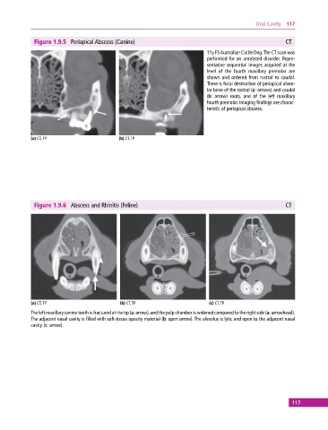
Figure 1.9.5 Periapical Abscess (Canine) CT
11y FS Australian Cattle Dog. The CT scan was
performed for an unrelated disorder. Repre
sentative sequential images acquired at the
level of the fourth maxillary premolar are
shown and ordered from rostral to caudal.
There is focal destruction of periapical alveo
lar bone of the rostral (a: arrows) and caudal
(b: arrow) roots, and of the left maxillary
fourth premolar. Imaging findings are charac
teristic of periapical abscess.
(a) CT, TP (b) CT, TP
Figure 1.9.6 Abscess and Rhinitis (Feline) CT
(a) CT, TP (b) CT, TP (c) CT, TP
The left maxillary canine tooth is fractured at the tip (a: arrow), and the pulp chamber is widened compared to the right side (a: arrowhead).
The adjacent nasal cavity is filled with soft‐tissue opacity material (b: open arrow). The alveolus is lytic and open to the adjacent nasal
cavity (c: arrow).
117