
Page 129 - Atlas of Small Animal CT and MRI
P. 129
Oral Cavity 119
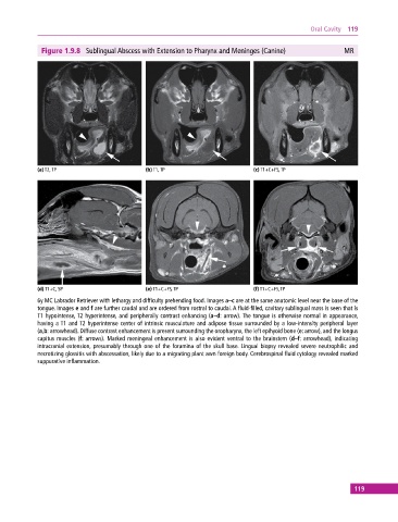
Figure 1.9.8 Sublingual Abscess with Extension to Pharynx and Meninges (Canine) MR
(a) T2, TP (b) T1, TP (c) T1+C+FS, TP
(d) T1+C, SP (e) T1+C+FS, TP (f) T1+C+FS, TP
6y MC Labrador Retriever with lethargy and difficulty prehending food. Images a–c are at the same anatomic level near the base of the
tongue. Images e and f are further caudal and are ordered from rostral to caudal. A fluid‐filled, cavitary sublingual mass is seen that is
T1 hypointense, T2 hyperintense, and peripherally contrast enhancing (a–d: arrow). The tongue is otherwise normal in appearance,
having a T1 and T2 hyperintense center of intrinsic musculature and adipose tissue surrounded by a low‐intensity peripheral layer
(a,b: arrowhead). Diffuse contrast enhancement is present surrounding the oropharynx, the left epihyoid bone (e: arrow), and the longus
capitus muscles (f: arrows). Marked meningeal enhancement is also evident ventral to the brainstem (d–f: arrowhead), indicating
intracranial extension, presumably through one of the foramina of the skull base. Lingual biopsy revealed severe neutrophilic and
necrotizing glossitis with abscessation, likely due to a migrating plant awn foreign body. Cerebrospinal fluid cytology revealed marked
suppurative inflammation.
119