Page 128 - Atlas of Small Animal CT and MRI
P. 128

118  Atlas of Small Animal CT and MRI


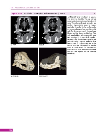
              Figure 1.9.7  Mandibular Osteomyelitis and Osteonecrosis (Canine)                            CT
                                                                              4y M Scottish Terrier with history of suppura­
                                                                              tive, ulcerative stomatitis.  The dog has had
                                                                              numerous tooth extractions performed previ­
                                                                              ously.  The molars and caudal premolars are
                                                                              missing. Representative sequential images
                                                                              acquired at the level of the first maxillary molar
                                                                              are shown and ordered from rostral to caudal
                                                                              (a,b). The alveolar processes in the maxilla are
                                                                              blunted, and the alveolar cavities have filled
                                                                              with organized bone (a,b: arrowheads). Large
                                                                              post‐extraction defects remain in the mandible,
                                                                              accompanied by alveolar bone destruction and
                                                                              adjacent  reactive  periosteal  production  (a,b:
                                                                              large arrows).  A focal gas collection is also
             (a) CT, TP                       (b) CT, TP                        evident within the right mandibular alveolar
                                                                              cavity (a:  small arrow).  The 3D renderings
                                                                              reveal the full extent of mandibular periodontal
                                                                              osteolysis  and  adjacent  reactive  periosteal
                                                                              remodeling.


             (c) CT, 3D, OBL                  (d) CT, 3D, LLAT


           118
   123   124   125   126   127   128   129   130   131   132   133