
Page 125 - Atlas of Small Animal CT and MRI
P. 125
Oral Cavity 115
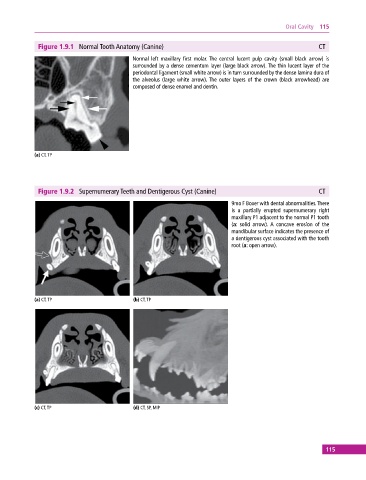
Figure 1.9.1 Normal Tooth Anatomy (Canine) CT
Normal left maxillary first molar. The central lucent pulp cavity (small black arrow) is
surrounded by a dense cementum layer (large black arrow). The thin lucent layer of the
periodontal ligament (small white arrow) is in turn surrounded by the dense lamina dura of
the alveolus (large white arrow). The outer layers of the crown (black arrowhead) are
composed of dense enamel and dentin.
(a) CT, TP
Figure 1.9.2 Supernumerary Teeth and Dentigerous Cyst (Canine) CT
9mo F Boxer with dental abnormalities. There
is a partially erupted supernumerary right
maxillary P1 adjacent to the normal P1 tooth
(a: solid arrow). A concave erosion of the
mandibular surface indicates the presence of
a dentigerous cyst associated with the tooth
root (a: open arrow).
(a) CT, TP (b) CT, TP
(c) CT, TP (d) CT, SP, MIP
115