
Page 120 - Atlas of Small Animal CT and MRI
P. 120
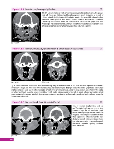
Figure 1.8.5  Reactive Lymphadenopathy (Canine)                                              CT

                                              12y MC Labrador Retriever with cervical necrotizing cellulitis and septicemia. The pharyn-
                                              geal soft tissues are thickened and fascial margins are poorly delineated as a result of
                                                diffuse regional cellulitis (asterisks). Mandibular lymph nodes are variably enlarged and are
                                                somewhat more spherical than normal (arrows). Contrast enhancement is uniform.
                                              Low‐density foci in some nodes are attributable to fat within the lymph node hilus.
                                              Microscopic evaluation of mandibular lymph nodes following euthanasia revealed marked
                                              diffuse plasmacytosis and lymphocytosis, consistent with node reactivity.


             (a) CT+C, TP


              Figure 1.8.6  Pyogranulomatous Lymphadenopathy & Lymph Node Abscess (Canine)                 CT


             (a) CT+C, TP                     (b) CT+C, TP
             7y MC Weimaraner with recent‐onset difficulty swallowing and pain on manipulation of the head and neck. Representative contrast‐
             enhanced CT images are at the level of the mandibular (a) and retropharyngeal (b) lymph nodes. Mandibular lymph nodes are enlarged
             and have extensive nodal and ill‐defined perinodal contrast enhancement (a: arrows). Similar findings are seen associated with the medial
             retropharyngeal lymph nodes (b: arrows). In addition, the left medial retropharyngeal lymph node is greatly enlarged and contains a fluid
               component ventrally consistent with frank abscessation. Aspiration cytology from the medial retropharyngeal lymph node confirmed suppurative
             inflammation and necrosis.


              Figure 1.8.7  Regional Lymph Node Metastasis (Canine)                                        CT

                                                                              10mo  F  German  Shepherd  Dog  with  an
                                                                                undifferentiated oral sarcoma, present since
                                                                              8  weeks of age. The left mandibular lymph
                                                                              node is markedly enlarged on the unenhanced
                                                                              image (a). Following contrast administration,
                                                                              there is peripheral enhancement of the man-
                                                                              dibular lymph node with a central nonenhanc-
                                                                              ing region representing metastasis (b: arrow).
                                                                              Fine‐needle aspiration cytology confirmed
                                                                              nodal metastasis.


             (a) CT, TP                       (b) CT+C, TP
           110