
Page 116 - Atlas of Small Animal CT and MRI
P. 116
106 Atlas of Small Animal CT and MRI
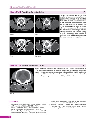
Figure 1.7.14 Parotid Duct Obstruction (Feline) CT
16y Domestic Longhair with bilateral facial
swelling. Representative unenhanced and com-
parable contrast‐enhanced images are ordered
from rostral to caudal. Bilateral well‐circum-
scribed, thin‐walled, fluid‐attenuating masses
are present ventrolaterally. Other images (not
shown) documented these masses are tubular.
Although the masses peripherally enhance, cen-
tral attenuation remains unchanged, indicative
of compartmentalized fluid. Aspiration cytology
confirmed the fluid was saliva. Attempts to
(a) CT, TP (b) CT, TP
catheterize the parotid ducts were unsuccessful
because of stenosis at the oral papilla.
(c) CT+C, TP (d) CT+C, TP
Figure 1.7.15 Sialocele with Sialoliths (Canine) CT
10y MC Maltese with a fluctuant ventral cervical mass. The CT image is at a level just rostral
to the mandibular salivary gland. An ill‐defined and diffusely hypoattenuating mass (arrow)
is present adjacent to the right external ear canal and tympanic bulla. Multiple focal mineral
opacities are distributed within the mass. Smaller numbers are seen on the left side. The
mass was thought to represent a sialocele containing multiple sialoliths.
(a) CT, TP
References findings in dogs with zygomatic sialadenitis: 11 cases (1990–2009).
J Am Vet Med Assoc. 2011 ed. 2011;239:1211–1218.
1. Weidner S, Probst A, Kneissl S. MR anatomy of salivary glands in 4. Watanabe K, Miyawaki S, Kanayama M, et al. First case of salivary
the dog. Anatom Histol Embryol. 2012;41:149–153. mucocele originating from the minor salivary gland of the soft
2. Kneissl S, Weidner S, Probst A. CT sialography in the dog – a palate in a dog. J Vet Med Sci. 2012;74:71–74.
cadaver study. Anatom Histol Embryol. 2011;40:397–401.
3. Cannon MS, Paglia D, Zwingenberger AL, Boroffka SA,
Hollingsworth SR, Wisner ER. Clinical and diagnostic imaging
106