
Page 113 - Atlas of Small Animal CT and MRI
P. 113
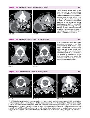
Figure 1.7.8  Mandibular Salivary Gland Abscess (Canine)                                    CT

                                                                             12y MC Rottweiler with a ventral cervical
                                                                             mass.  Representative  contrast‐enhanced
                                                                             images are ordered from rostral to caudal.
                                                                             There is a large predominantly fluid attenuat-
                                                                             ing cavitary mass contiguous with the lateral
                                                                             margin of the right mandibular salivary gland
                                                                             (a:  asterisk). The lateral contour of the gland is
                                                                             altered, and the fluid center extends into the
                                                                             glandular parenchyma (a). The mass is thick
                                                                             walled,  peripherally enhancing,  and  poorly
                                                                             margined. The center consists predominantly
                                                                             of fluid but also includes a small volume of
                                                                             fragmented gas (b). A mixed bacterial popula-
                                                                             tion was cultured from a fluid aspirate.
            (a) CT+C, TP                     (b) CT+C, TP


              Figure 1.7.9  Mandibular Salivary Adenocarcinoma (Feline)                                   CT

                                                                             8y FS  Siamese  with a ventral cervical mass.
                                                                             Representative images are at the level of the
                                                                             mandibular salivary glands.  There is a large,
                                                                             spherical, low‐attenuation mandibular salivary
                                                                             mass in the right ventral cervical region
                                                                             (a,b: large arrow). The mass contrast enhances
                                                                             nonuniformly centrally and has a thin but prom-
                                                                             inent peripheral rim of enhancement (b). The
                                                                             normal left mandibular salivary gland is also
                                                                             evident (a,b: small arrow). Biopsy confirmed
                                                                             mandibular salivary adenocarcinoma.


            (a) CT, TP                       (b) CT+C, TP


              Figure 1.7.10  Parotid Salivary Adenocarcinoma (Canine)                                     CT


            (a) CT, TP                       (b) CT+C, TP                     (c) CT+C, TP
            13y MC Golden Retriever with a slowly enlarging mass. There is a large, irregularly margined mass arising from the right parotid salivary
            gland (a,b: large arrow). The normal left parotid gland is also seen (a,b: small arrow). The cranial poles of the mandibular salivary
            glands can also be seen medial to the parotid glands (a,b: arrowheads). An enlarged right mandibular lymph node is also present
            (a,b: adjacent to left arrowhead). The mass nonuniformly contrast enhances revealing a central cavitary component (b). Further caudally,
            a markedly enlarged right medial retropharyngeal lymph node is evident (c: arrow) adjacent to the mandibular gland (c: arrowhead).
            Biopsy of the mass and lymph nodes confirmed a diagnosis of parotid salivary adenocarcinoma with regional lymph node metastasis.