
Page 130 - Atlas of Small Animal CT and MRI
P. 130
120 Atlas of Small Animal CT and MRI
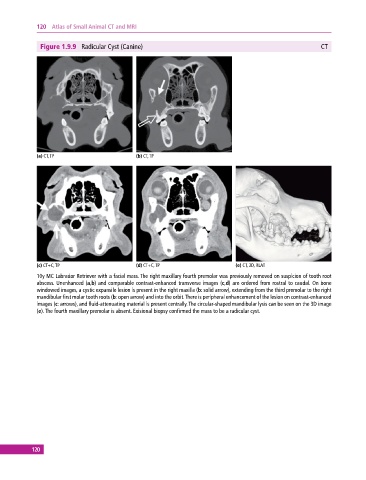
Figure 1.9.9 Radicular Cyst (Canine) CT
(a) CT, TP (b) CT, TP
(c) CT+C, TP (d) CT+C, TP (e) CT, 3D, RLAT
10y MC Labrador Retriever with a facial mass. The right maxillary fourth premolar was previously removed on suspicion of tooth root
abscess. Unenhanced (a,b) and comparable contrastenhanced transverse images (c,d) are ordered from rostral to caudal. On bone
windowed images, a cystic expansile lesion is present in the right maxilla (b: solid arrow), extending from the third premolar to the right
mandibular first molar tooth roots (b: open arrow) and into the orbit. There is peripheral enhancement of the lesion on contrastenhanced
images (c: arrows), and fluid‐attenuating material is present centrally. The circular‐shaped mandibular lysis can be seen on the 3D image
(e). The fourth maxillary premolar is absent. Exisional biopsy confirmed the mass to be a radicular cyst.
120