
Page 135 - Atlas of Small Animal CT and MRI
P. 135
Oral Cavity 125
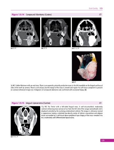
Figure 1.9.14 Compound Odontoma (Canine) CT
(a) CT, TP (b) CT, TP (c) CT+C, TP
(d) GP
3y MC Golden Retriever with an oral mass. There is an expansile, primarily productive mass in the left mandible on the lingual and buccal
sides of the teeth (a: arrows). There is cortical lysis, but the margin of the mass is smooth and regular. No soft‐tissue component is present
on contrast‐enhanced images (c). A diagnosis of compound odontoma was confirmed with excisional biopsy (d).
Figure 1.9.15 Lingual Liposarcoma (Canine) CT
12y MC Fox Terrier with a left‐sided lingual mass. A well‐circumscribed, moderately
contrast‐enhancing mass (arrow) arises from the left side of the tongue (arrowhead) and is
displaced ventrally by the overlying endotracheal catheter. The tongue is otherwise normal
in appearance, having a relatively low‐density center of intrinsic musculature and adipose
tissue surrounded by a soft‐tissue dense peripheral layer. Biopsy of the mass revealed it to
be a moderately well‐differentiated liposarcoma.
(a) CT+C, TP
125