
Page 137 - Atlas of Small Animal CT and MRI
P. 137
Oral Cavity 127
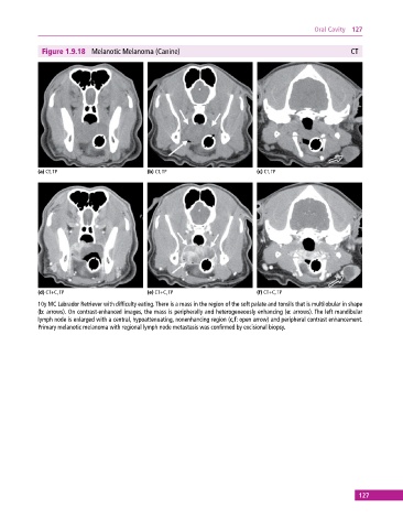
Figure 1.9.18 Melanotic Melanoma (Canine) CT
(a) CT, TP (b) CT, TP (c) CT, TP
(d) CT+C, TP (e) CT+C, TP (f) CT+C, TP
10y MC Labrador Retriever with difficulty eating. There is a mass in the region of the soft palate and tonsils that is multilobular in shape
(b: arrows). On contrast‐enhanced images, the mass is peripherally and heterogeneously enhancing (e: arrows). The left mandibular
lymph node is enlarged with a central, hypoattenuating, nonenhancing region (c,f: open arrow) and peripheral contrast enhancement.
Primary melanotic melanoma with regional lymph node metastasis was confirmed by excisional biopsy.
127