
Page 136 - Atlas of Small Animal CT and MRI
P. 136
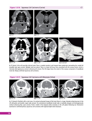
Figure 1.9.16  Squamous Cell Carcinoma (Canine)                                              CT


             (a) CT, TP                       (b) CT, TP


             (c) CT+C, TP                     (d) CT+C, TP                     (e) CT, 3D, RLAT
             8y FS Border Collie with possible infected tooth. There is marked osteolysis and irregular bone production surrounding the caudal left
             mandible (a,b: open arrows). Multiple teeth are absent. There is a large soft‐tissue mass associated with the osseous lesion, which is
             intensely, heterogeneously contrast enhancing (d: arrows). The 3D image shows the extent of the osseous component of mandibular
             lesion (e). Biopsy confirmed squamous cell carcinoma.


              Figure 1.9.17  Squamous Cell Carcinoma with Metastasis (Feline)                              CT


             (a) CT+C, TP                     (b) CT+C, TP                     (c) CT+C, TP
             9y FS Domestic Shorthair with a neck mass. On contrast‐enhanced images of the head, there is a large, intensely enhancing mass in the
             left soft palate and tonsillar region (a,b: arrows). The contralateral mandibular lymph node is markedly enlarged and heterogeneously
             contrast enhancing (b,c: open arrows). Fluid is visible in the tympanic bulla secondary to the soft palate thickening (c). Postmortem
             examination confirmed primary squamous cell carcinoma with regional lymph node metastasis.


           126