
Page 133 - Atlas of Small Animal CT and MRI
P. 133
Oral Cavity 123
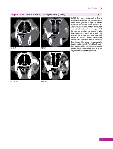
Figure 1.9.12 Amyloid Producing Odontogenic Tumor (Canine) CT
8y M Terrier mix with orbital swelling. There is
an expansile productive and destructive bone
lesion involving the rostral aspect of the left
zygomatic arch and left caudal maxilla (a,b).
After intravenous administration of iodinated
contrast medium, the soft‐tissue component of
the lesion has a multicameral appearance with
hypoattenuating loculated regions and good
enhancement of interspersed soft tissues and
septae (c: arrows). Contrast enhancement
demonstrates extension of the mass proximally
from the level of the second premolars caudally
into the ventral orbit (d). The left fourth maxil
lary premolar and left maxillary molars are not
(a) CT, TP (b) CT, TP present. Biopsy confirmed the mass to be an
amyloid‐producing odontogenic tumor.
(c) CT+C, TP (d) CT+C, TP
123