Page 134 - Atlas of Small Animal CT and MRI
P. 134

124  Atlas of Small Animal CT and MRI


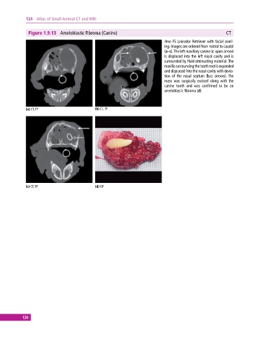
              Figure 1.9.13  Ameloblastic Fibroma (Canine)                                                 CT
                                                                              4mo FS Labrador Retriever with facial swell­
                                                                              ing. Images are ordered from rostral to caudal
                                                                              (a–c). The left maxillary canine (c: open arrow)
                                                                              is displaced into the left nasal cavity and is
                                                                              surrounded by fluid‐attenuating material. The
                                                                              maxilla surrounding the tooth root is expanded
                                                                              and displaced into the nasal cavity with devia­
                                                                              tion of the nasal septum (b,c:  arrows). The
                                                                              mass  was  surgically  excised  along  with  the
                                                                              canine tooth and was confirmed to be an
                                                                              ameloblastic fibroma (d).


             (a) CT, TP                       (b) CT, TP


             (c) CT, TP                       (d) GP


           124
   129   130   131   132   133   134   135   136   137   138   139