
Page 138 - Atlas of Small Animal CT and MRI
P. 138
128 Atlas of Small Animal CT and MRI
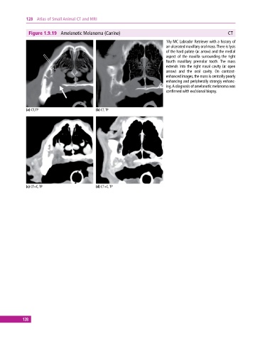
Figure 1.9.19 Amelanotic Melanoma (Canine) CT
10y MC Labrador Retriever with a history of
an ulcerated maxillary oral mass. There is lysis
of the hard palate (a: arrow) and the medial
aspect of the maxilla surrounding the right
fourth maxillary premolar tooth. The mass
extends into the right nasal cavity (a: open
arrow) and the oral cavity. On contrast‐
enhanced images, the mass is centrally poorly
enhancing and peripherally strongly enhanc
ing. A diagnosis of amelanotic melanoma was
confirmed with excisional biopsy.
(a) CT, TP (b) CT, TP
(c) CT+C, TP (d) CT+C, TP
128