
Page 100 - Atlas of Small Animal CT and MRI
P. 100
90 Atlas of Small Animal CT and MRI
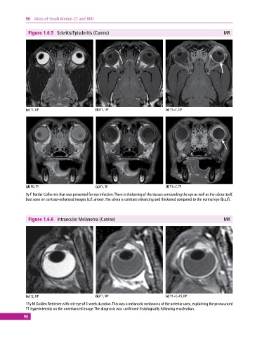
Figure 1.6.5 Scleritis/Episcleritis (Canine) MR
(a) T2, DP (b) T1, DP (c) T1+C, DP
(d) PD, TP (e) T1, TP (f) T1+C, TP
9y F Border Collie mix that was presented for eye infection. There is thickening of the tissues surrounding the eye as well as the sclera itself,
best seen on contrast‐enhanced images (c,f: arrow). The sclera is contrast enhancing and thickened compared to the normal eye (b,c,f).
Figure 1.6.6 Intraocular Melanoma (Canine) MR
(a) T2, DP (b) T1, DP (c) T1+C+FS, DP
11y M Golden Retriever with red‐eye of 3-week duration. This was a melanotic melanoma of the anterior uvea, explaining the pronounced
T1 hyperintensity on the unenhanced image. The diagnosis was confirmed histologically following enucleation.
90