
Page 75 - Atlas of Small Animal CT and MRI
P. 75
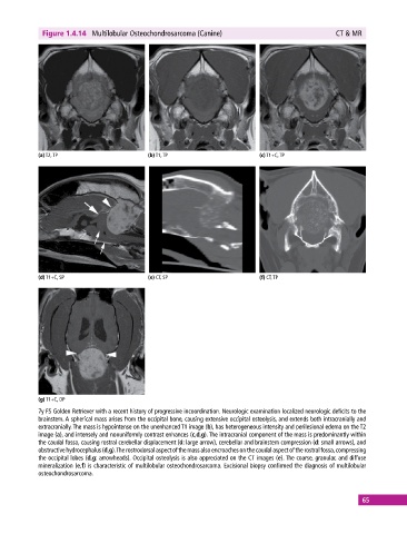
Figure 1.4.14  Multilobular Osteochondrosarcoma (Canine)                               CT & MR


            (a) T2, TP                       (b) T1, TP                       (c) T1+C, TP


            (d) T1+C, SP                     (e) CT, SP                       (f) CT, TP


            (g) T1+C, DP
            7y FS Golden Retriever with a recent history of progressive incoordination. Neurologic examination localized neurologic deficits to the
            brainstem. A spherical mass arises from the occipital bone, causing extensive occipital osteolysis, and extends both intracranially and
            extracranially. The mass is hypointense on the unenhanced T1 image (b), has heterogeneous intensity and perilesional edema on the T2
            image (a), and intensely and nonuniformly contrast enhances (c,d,g). The intracranial component of the mass is predominantly within
            the caudal fossa, causing rostral cerebellar displacement (d: large arrow), cerebellar and brainstem compression (d: small arrows), and
            obstructive hydrocephalus (d,g). The rostrodorsal aspect of the mass also encroaches on the caudal aspect of the rostral fossa,  compressing
            the occipital lobes (d,g: arrowheads). Occipital osteolysis is also appreciated on the CT images (e). The coarse, granular, and diffuse
            mineralization (e,f) is characteristic of multilobular osteochondrosarcoma. Excisional biopsy confirmed the diagnosis of multilobular
            osteochondrosarcoma.


                                                                                                             65