
Page 63 - Atlas of Small Animal CT and MRI
P. 63
Temporomandibular Joint 53
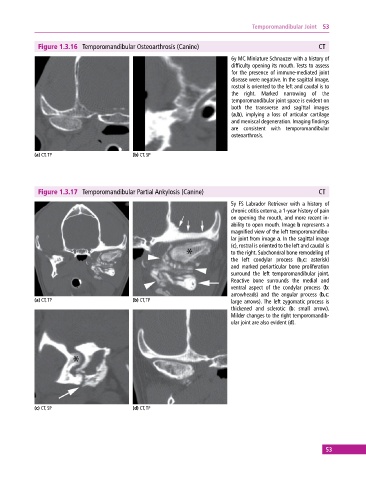
Figure 1.3.16 Temporomandibular Osteoarthrosis (Canine) CT
6y MC Miniature Schnauzer with a history of
difficulty opening its mouth. Tests to assess
for the presence of immune‐mediated joint
disease were negative. In the sagittal image,
rostral is oriented to the left and caudal is to
the right. Marked narrowing of the
temporomandibular joint space is evident on
both the transverse and sagittal images
(a,b), implying a loss of articular cartilage
and meniscal degeneration. Imaging findings
are consistent with temporomandibular
osteoarthrosis.
(a) CT, TP (b) CT, SP
Figure 1.3.17 Temporomandibular Partial Ankylosis (Canine) CT
5y FS Labrador Retriever with a history of
chronic otitis externa, a 1‐year history of pain
on opening the mouth, and more recent in-
ability to open mouth. Image b represents a
magnified view of the left temporomandibu-
lar joint from image a. In the sagittal image
(c), rostral is oriented to the left and caudal is
to the right. Subchondral bone remodeling of
the left condylar process (b,c: asterisk)
and marked periarticular bone proliferation
surround the left temporomandibular joint.
Reactive bone surrounds the medial and
ventral aspect of the condylar process (b:
arrowheads) and the angular process (b,c:
(a) CT, TP (b) CT, TP large arrows). The left zygomatic process is
thickened and sclerotic (b: small arrow).
Milder changes to the right temporomandib-
ular joint are also evident (d).
(c) CT, SP (d) CT, TP
53