
Page 58 - Atlas of Small Animal CT and MRI
P. 58
48 Atlas of Small Animal CT and MRI
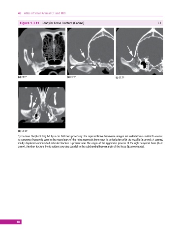
Figure 1.3.11 Condylar Fossa Fracture (Canine) CT
(a) CT, TP (b) CT, TP (c) CT, TP
(d) CT, DP
1y German Shepherd Dog hit by a car 24 hours previously. The representative transverse images are ordered from rostral to caudal.
A transverse fracture is seen in the rostral part of the right zygomatic bone near its articulation with the maxilla (a: arrow). A second,
mildly displaced comminuted articular fracture is present near the origin of the zygomatic process of the right temporal bone (b–d:
arrow). Another fracture line is evident coursing parallel to the subchondral bone margin of the fossa (b: arrowheads).
48