
Page 53 - Atlas of Small Animal CT and MRI
P. 53
Temporomandibular Joint 43
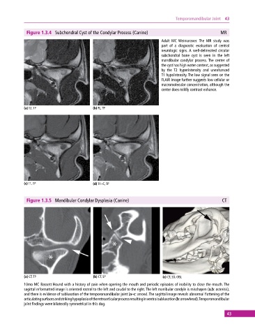
Figure 1.3.4 Subchondral Cyst of the Condylar Process (Canine) MR
Adult MC Weimaraner. The MR study was
part of a diagnostic evaluation of central
neurologic signs. A well‐delineated circular
subchondral bone cyst is seen in the left
mandibular condylar process. The center of
the cyst has high water content, as suggested
by the T2 hyperintensity and unenhanced
T1 hypointensity. The low signal seen on the
FLAIR image further suggests low cellular or
macromolecular concentration, although the
center does mildly contrast enhance.
(a) T2, TP (b) FL, TP
(c) T1, TP (d) T1+C, TP
Figure 1.3.5 Mandibular Condylar Dysplasia (Canine) CT
(a) CT, TP (b) CT, SP (c) CT, 3D, OBL
10mo MC Bassett Hound with a history of pain when opening the mouth and periodic episodes of inability to close the mouth. The
sagittal reformatted image is oriented rostral to the left and caudal to the right. The left manibular condyle is misshapen (a,b: asterisk),
and there is evidence of subluxation of the temporomandibular joint (a–c: arrow). The sagittal image reveals abnormal flattening of the
articulating surfaces and striking hypoplasia of the retroarticular process resulting in ventral subluxation (b: arrowhead). Temporomandibular
joint findings were bilaterally symmetrical in this dog.
43