Page 55 - Atlas of Small Animal CT and MRI
P. 55

Temporomandibular Joint  45


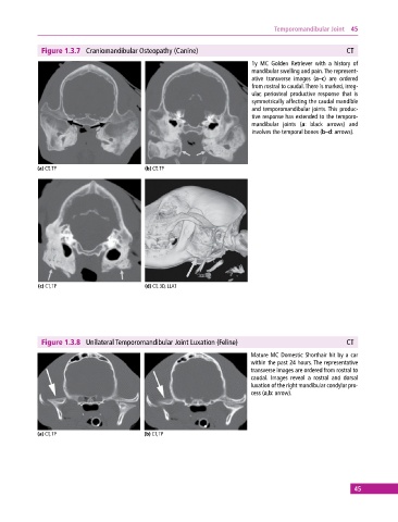
              Figure 1.3.7  Craniomandibular Osteopathy (Canine)                                          CT
                                                                             1y MC Golden Retriever with a history of
                                                                             mandibular swelling and pain. The represent-
                                                                             ative transverse images (a–c) are ordered
                                                                             from rostral to caudal. There is marked, irreg-
                                                                             ular, periosteal productive response that is
                                                                             symmetrically affecting the caudal mandible
                                                                             and  temporomandibular joints. This produc-
                                                                             tive response has extended to the temporo-
                                                                             mandibular joints (a: black arrows) and
                                                                             involves the temporal bones (b–d: arrows).


            (a) CT, TP                       (b) CT, TP


             (c) CT, TP                      (d) CT, 3D, LLAT


              Figure 1.3.8  Unilateral Temporomandibular Joint Luxation (Feline)                          CT
                                                                             Mature MC Domestic Shorthair hit by a car
                                                                             within the past 24 hours. The representative
                                                                             transverse images are ordered from rostral to
                                                                             caudal. Images reveal a rostral and dorsal
                                                                             luxation of the right mandibular condylar pro-
                                                                             cess (a,b: arrow).


            (a) CT, TP                       (b) CT, TP


                                                                                                             45
   50   51   52   53   54   55   56   57   58   59   60