
Page 52 - Atlas of Small Animal CT and MRI
P. 52
42 Atlas of Small Animal CT and MRI
Figure 1.3.2 Normal Temporomandibular Joint (Canine) MR
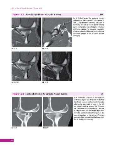
2y M Pit Bull Terrier. The condyloid process
and region of the mandibular fossa appear T1
and T2 hyperintense centrally because of
medullary fat, with a well to poorly defined
signal void peripherally defining the subchon-
dral bone margins. The apparent irregularity
of the subchondral bone of the condyle on
transverse images is due to partial volume
averaging.
(a) T1, TP (b) T1, SP
(c) T1+C, TP (d) T2, TP
Figure 1.3.3 Subchondral Cyst of the Condylar Process (Canine) CT
3y M Rottweiler. A CT scan of the head was
performed as part of a diagnostic evaluation
for chronic otitis. A well‐delineated circular
subchondral bone cyst is seen in the left
mandibular condylar process (a). Contents
are fluid‐dense and surrounded by a thin rim
of compact bone. The left condyle (b) is unre-
markable and included in this figure in the
same orientation for comparison. The cyst
was clinically silent and identified as an inci-
dental finding on this study.
(a) CT, TP (b) CT, TP
42