Page 56 - Atlas of Small Animal CT and MRI
P. 56

46  Atlas of Small Animal CT and MRI


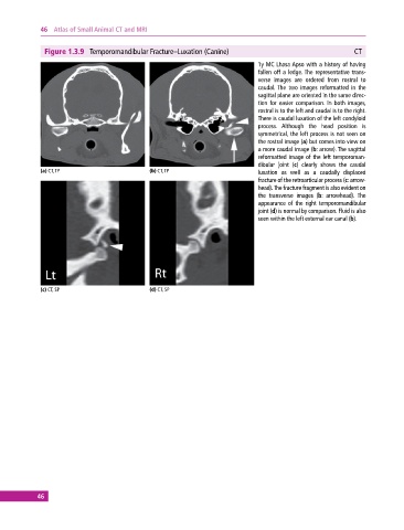
              Figure 1.3.9  Temporomandibular Fracture–Luxation (Canine)                                   CT
                                                                              1y MC Lhasa Apso with a history of having
                                                                              fallen off a ledge. The representative trans-
                                                                              verse images are ordered from rostral to
                                                                                caudal. The two images reformatted in the
                                                                              sagittal plane are oriented in the same direc-
                                                                              tion for easier comparison. In both images,
                                                                              rostral is to the left and caudal is to the right.
                                                                              There is caudal luxation of the left condyloid
                                                                              process.  Although the head position is
                                                                                symmetrical, the left process is not seen on
                                                                              the rostral image (a) but comes into view on
                                                                              a more caudal image (b: arrow). The sagittal
                                                                              reformatted image of the left temporoman-
                                                                              dibular joint (c) clearly shows the caudal
             (a) CT, TP                       (b) CT, TP                        luxation  as  well  as  a  caudally  displaced
                                                                                fracture of the retroarticular process (c: arrow-
                                                                              head). The fracture fragment is also evident on
                                                                              the  transverse  images  (b:  arrowhead).  The
                                                                              appearance of the right temporomandibular
                                                                              joint (d) is normal by comparison. Fluid is also
                                                                              seen within the left external ear canal (b).


             (c) CT, SP                       (d) CT, SP


            46
   51   52   53   54   55   56   57   58   59   60   61