
Page 32 - Atlas of Small Animal CT and MRI
P. 32
22 Atlas of Small Animal CT and MRI
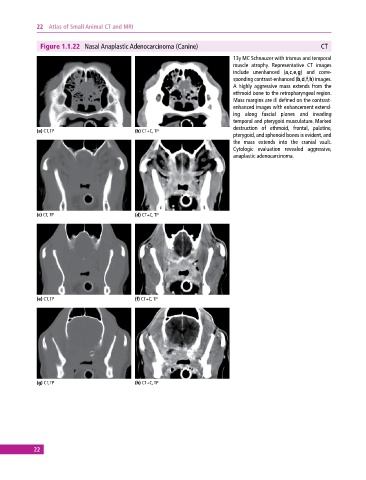
Figure 1.1.22 Nasal Anaplastic Adenocarcinoma (Canine) CT
13y MC Schnauzer with trismus and temporal
muscle atrophy. Representative CT images
include unenhanced (a,c,e,g) and corre-
sponding contrast‐enhanced (b,d,f,h) images.
A highly aggressive mass extends from the
ethmoid bone to the retropharyngeal region.
Mass margins are ill defined on the contrast‐
enhanced images with enhancement extend-
ing along fascial planes and invading
temporal and pterygoid musculature. Marked
destruction of ethmoid, frontal, palatine,
(a) CT, TP (b) CT+C, TP
pterygoid, and sphenoid bones is evident, and
the mass extends into the cranial vault.
Cytologic evaluation revealed aggressive,
anaplastic adenocarcinoma.
(c) CT, TP (d) CT+C, TP
(e) CT, TP (f) CT+C, TP
(g) CT, TP (h) CT+C, TP
22