
Page 29 - Atlas of Small Animal CT and MRI
P. 29
Nasal Cavity and Paranasal Sinuses 19
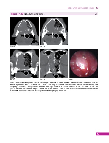
Figure 1.1.19 Nasal Lymphoma (Canine) CT
(a) CT, TP (b) CT, TP
(c) CT, TP (d) CT, TP (e) ES
3y MC Rhodesian Ridgeback with a 3‐month history of nasal discharge and stertor. There is a predominantly right‐sided nasal mass that
extends beyond midline to fill the ventral part of the left nasal cavity rostral to the maxillary sinuses. The mass extends caudally to the
nasopharynx (d: asterisk). Nearly complete osteolysis of the right nasal ectoturbinates is evident (a,b), and there is destruction of the
palatine portion of the maxilla and the palatine bone (a,b: arrow). Vomer bone destruction is also present where the mass extends across
midline (a,b: arrowhead). Retrograde rhinoscopy revealed a nasopharyngeal mass (e).
19