
Page 24 - Atlas of Small Animal CT and MRI
P. 24
14 Atlas of Small Animal CT and MRI
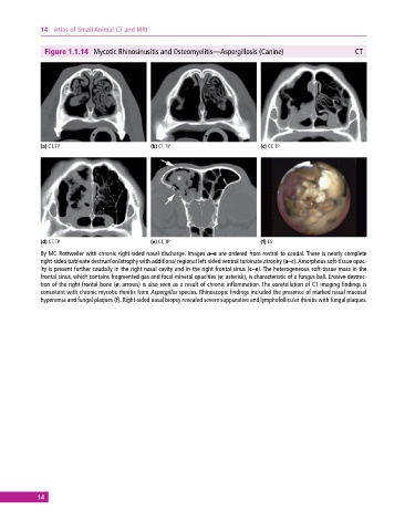
Figure 1.1.14 Mycotic Rhinosinusitis and Osteomyelitis—Aspergillosis (Canine) CT
(a) CT, TP (b) CT, TP (c) CT, TP
(d) CT, TP (e) CT, TP (f) ES
8y MC Rottweiler with chronic right‐sided nasal discharge. Images a–e are ordered from rostral to caudal. There is nearly complete
right‐sided turbinate destruction/atrophy with additional regional left‐sided ventral turbinate atrophy (a–c). Amorphous soft‐tissue opac-
ity is present further caudally in the right nasal cavity and in the right frontal sinus (c–e). The heterogeneous soft‐tissue mass in the
frontal sinus, which contains fragmented gas and focal mineral opacities (e: asterisk), is characteristic of a fungus ball. Erosive destruc-
tion of the right frontal bone (e: arrows) is also seen as a result of chronic inflammation. The constellation of CT imaging findings is
consistent with chronic mycotic rhinitis from Aspergillus species. Rhinoscopic findings included the presence of marked nasal mucosal
hyperemia and fungal plaques (f). Right‐sided nasal biopsy revealed severe suppurative and lymphofollicular rhinitis with fungal plaques.
14