
Page 23 - Atlas of Small Animal CT and MRI
P. 23
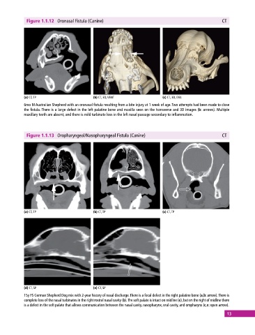
Figure 1.1.12  Oronasal Fistula (Canine)                                                    CT


            (a) CT, TP                       (b) CT, 3D, VENT                 (c) CT, 3D, OBL
            6mo M Australian Shepherd with an oronasal fistula resulting from a bite injury at 1 week of age. Two attempts had been made to close
            the fistula. There is a large defect in the left palatine bone and maxilla seen on the transverse and 3D images (b: arrows). Multiple
              maxillary teeth are absent, and there is mild turbinate loss in the left nasal passage secondary to inflammation.


              Figure 1.1.13  Oropharyngeal/Nasopharyngeal Fistula (Canine)                                CT


            (a) CT, TP                       (b) CT, TP                       (c) CT, TP


            (d) CT, SP                       (e) CT, SP
            15y FS German Shepherd Dog mix with 2‐year history of nasal discharge. There is a focal defect in the right palatine bone (a,b: arrow). There is
            complete loss of the nasal turbinates in the right rostral nasal cavity (b). The soft palate is intact on midline (e), but on the right of midline there
            is a defect in the soft palate that allows communication between the nasal cavity, nasopharynx, oral cavity, and oropharynx (c,e: open arrow).

                                                                                                             13