
Page 20 - Atlas of Small Animal CT and MRI
P. 20
10 Atlas of Small Animal CT and MRI
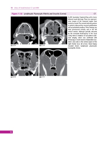
Figure 1.1.8 Lymphocytic Plasmacytic Rhinitis and Sinusitis (Canine) CT
9y MC Australian Shepherd Dog with chronic
bilateral nasal discharge. These are represent-
ative images of the sinonasal region from
rostral to caudal. The normal turbinate pattern
is partially obscured by mucosal proliferation
and accumulated exudates. These findings are
most pronounced rostrally and in the left
ventral meatus. Although partially obscured
by the increased fluid‐opacity in the nasal
cavity, there is evidence of nonuniform turbi-
nate atrophy, which was confirmed with
rhinoscopy. A small volume of dependent exu-
date is also seen in the ventral aspect of the
right frontal sinus (d: arrow). Nasal biopsy
(a) CT, TP (b) CT, TP revealed chronic lymphocytic plasmocytic
neutrophilic rhinitis.
(c) CT, TP (d) CT, TP
10