
Page 26 - Atlas of Small Animal CT and MRI
P. 26
16 Atlas of Small Animal CT and MRI
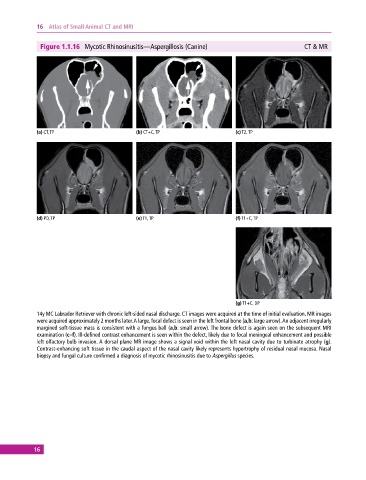
Figure 1.1.16 Mycotic Rhinosinusitis—Aspergillosis (Canine) CT & MR
(a) CT, TP (b) CT+C, TP (c) T2, TP
(d) PD, TP (e) T1, TP (f) T1+C, TP
(g) T1+C, DP
14y MC Labrador Retriever with chronic left‐sided nasal discharge. CT images were acquired at the time of initial evaluation. MR images
were acquired approximately 2 months later. A large, focal defect is seen in the left frontal bone (a,b: large arrow). An adjacent irregularly
margined soft‐tissue mass is consistent with a fungus ball (a,b: small arrow). The bone defect is again seen on the subsequent MRI
examination (c–f). Ill‐defined contrast enhancement is seen within the defect, likely due to focal meningeal enhancement and possible
left olfactory bulb invasion. A dorsal plane MR image shows a signal void within the left nasal cavity due to turbinate atrophy (g).
Contrast‐enhancing soft tissue in the caudal aspect of the nasal cavity likely represents hypertrophy of residual nasal mucosa. Nasal
biopsy and fungal culture confirmed a diagnosis of mycotic rhinosinusitis due to Aspergillus species.
16