
Page 197 - Atlas of Small Animal CT and MRI
P. 197
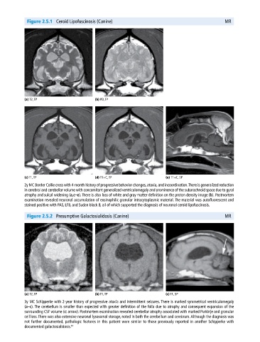
Figure 2.5.1  Ceroid Lipofuscinosis (Canine)                                               MR


            (a) T2, TP                       (b) PD, TP


            (c) T1, TP                       (d) T1+C, TP                     (e) T1+C, SP
            2y MC Border Collie cross with 4‐month history of progressive behavior changes, ataxia, and incoordination. There is generalized  reduction
            in cerebral and cerebellar volume with concomitant generalized ventriculomegaly and prominence of the subarachnoid space due to gyral
            atrophy and sulcal widening (a,c–e). There is also loss of white and gray matter definition on the proton density image (b). Postmortem
            examination revealed neuronal accumulation of eosinophilic granular intracytoplasmic material. The material was autofluorescent and
            stained positive with PAS, LFB, and Sudan black B, all of which supported the diagnosis of neuronal ceroid lipofuscinosis.


              Figure 2.5.2  Presumptive Galactosialidosis (Canine)                                       MR


            (a) T2, TP                       (b) T1, TP                       (c) T1, SP
            3y MC Schipperke with 2‐year history of progressive ataxia and intermittent seizures. There is marked symmetrical ventriculomegaly
            (a–c). The cerebellum is smaller than expected with greater definition of the folia due to atrophy and consequent expansion of the
              surrounding CSF volume (c: arrow). Postmortem examination revealed cerebellar atrophy associated with marked Purkinje and granular
            cell loss. There was also extensive neuronal lysosomal storage, noted in both the cerebellum and cerebrum. Although the diagnosis was
            not further documented, pathologic features in this patient were similar to those previously reported in another Schipperke with
              documented galactosialidosis. 14