
Page 192 - Atlas of Small Animal CT and MRI
P. 192
182 Atlas of Small Animal CT and MRI
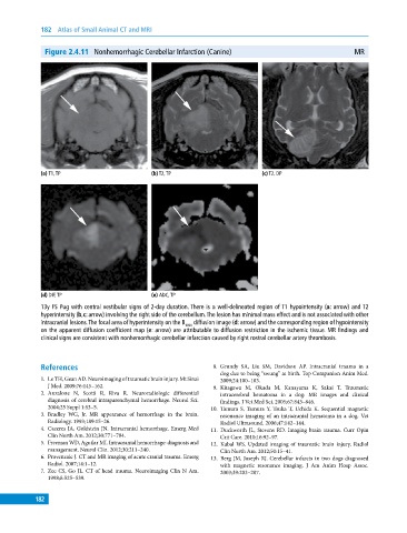
Figure 2.4.11 Nonhemorrhagic Cerebellar Infarction (Canine) MR
(a) T1, TP (b) T2, TP (c) T2, DP
(d) DIF, TP (e) ADC, TP
13y FS Pug with central vestibular signs of 2‐day duration. There is a well‐delineated region of T1 hypointensity (a: arrow) and T2
hyperintensity (b,c: arrow) involving the right side of the cerebellum. The lesion has minimal mass effect and is not associated with other
intracranial lesions. The focal area of hyperintensity on the B 1000 diffusion image (d: arrow) and the corresponding region of hypointensity
on the apparent diffusion coefficient map (e: arrow) are attributable to diffusion restriction in the ischemic tissue. MR findings and
clinical signs are consistent with nonhemorrhagic cerebellar infarction caused by right rostral cerebellar artery thrombosis.
References 8. Grundy SA, Liu SM, Davidson AP. Intracranial trauma in a
dog due to being “swung” at birth. Top Companion Anim Med.
1. Le TH, Gean AD. Neuroimaging of traumatic brain injury. Mt Sinai 2009;24:100–103.
J Med. 2009;76:145–162. 9. Kitagawa M, Okada M, Kanayama K, Sakai T. Traumatic
2. Anzalone N, Scotti R, Riva R. Neuroradiologic differential intracerebral hematoma in a dog: MR images and clinical
diagnosis of cerebral intraparenchymal hemorrhage. Neurol Sci. findings. J Vet Med Sci. 2005;67:843–846.
2004;25 Suppl 1:S3–5. 10. Tamura S, Tamura Y, Tsuka T, Uchida K. Sequential magnetic
3. Bradley WG, Jr. MR appearance of hemorrhage in the brain. resonance imaging of an intracranial hematoma in a dog. Vet
Radiology. 1993;189:15–26. Radiol Ultrasound. 2006;47:142–144.
4. Caceres JA, Goldstein JN. Intracranial hemorrhage. Emerg Med 11. Duckworth JL, Stevens RD. Imaging brain trauma. Curr Opin
Clin North Am. 2012;30:771–794. Crit Care. 2010;16:92–97.
5. Freeman WD, Aguilar MI. Intracranial hemorrhage: diagnosis and 12. Kubal WS. Updated imaging of traumatic brain injury. Radiol
management. Neurol Clin. 2012;30:211–240. Clin North Am. 2012;50:15–41.
6. Provenzale J. CT and MR imaging of acute cranial trauma. Emerg 13. Berg JM, Joseph RJ. Cerebellar infarcts in two dogs diagnosed
Radiol. 2007;14:1–12. with magnetic resonance imaging. J Am Anim Hosp Assoc.
7. Zee CS, Go JL. CT of head trauma. Neuroimaging Clin N Am. 2003;39:203–207.
1998;8:525–539.
182