
Page 191 - Atlas of Small Animal CT and MRI
P. 191
Trauma, Hemorrhage, and Vascular Disorders 181
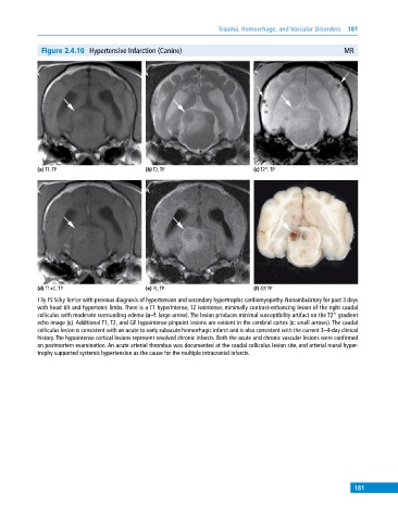
Figure 2.4.10 Hypertensive Infarction (Canine) MR
(a) T1, TP (b) T2, TP (c) T2*, TP
(d) T1+C, TP (e) FL, TP (f) GP, TP
13y FS Silky Terrier with previous diagnosis of hypertension and secondary hypertrophic cardiomyopathy. Nonambulatory for past 3 days
with head tilt and hypertonic limbs. There is a T1 hyperintense, T2 isointense, minimally contrast‐enhancing lesion of the right caudal
colliculus with moderate surrounding edema (a–f: large arrow). The lesion produces minimal susceptibility artifact on the T2* gradient
echo image (c). Additional T1, T2, and GE hypointense pinpoint lesions are evident in the cerebral cortex (c: small arrows). The caudal
colliculus lesion is consistent with an acute to early subacute hemorrhagic infarct and is also consistent with the current 3–4‐day clinical
history. The hypointense cortical lesions represent resolved chronic infarcts. Both the acute and chronic vascular lesions were confirmed
on postmortem examination. An acute arterial thrombus was documented at the caudal colliculus lesion site, and arterial mural hyper-
trophy supported systemic hypertension as the cause for the multiple intracranial infarcts.
181