
Page 190 - Atlas of Small Animal CT and MRI
P. 190
180 Atlas of Small Animal CT and MRI
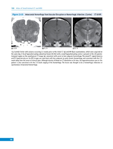
Figure 2.4.9 Intracranial Hemorrhage from Vascular Disruption or Hemorrhagic Infarction. (Canine) CT & MR
(a) CT, TP (b) T1, TP (c) T2, TP
12y Scottish Terrier with seizures occurring 2.5 weeks prior to the initial CT (a) and MR (b,c) examinations, which were acquired on
the same day. A focal hyperattenuating subcortical lesion (65 HU) with a small hypoattenuating center is present in the left parieto-
temporal region on the unenhanced CT image (a), consistent with acute to late subacute hemorrhage. The mixed T1 signal intensity
and T2 hypointensity on the MR images are consistent with late subacute to early chronic hemorrhage and consistent with the 2.5‐
week delay from the onset of clinical signs. Although density of blood on CT diminishes with time, the hyperattenuation seen in this
patient is also consistent with the 2.5‐week staging of the hemorrhage. The lesion was thought to be a hemorrhagic infarction or
spontaneous intracranial hemorrhage.
180