
Page 189 - Atlas of Small Animal CT and MRI
P. 189
Trauma, Hemorrhage, and Vascular Disorders 179
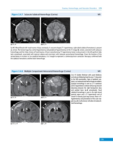
Figure 2.4.7 Subacute Subdural Hemorrhage (Canine) MR
(a) T1, TP (b) T2, TP
5y MC Mixed Breed with head trauma 4 days previously. A crescent‐shaped, T1 hyperintense, right‐sided subdural hematoma is present
(a: arrow). The hemorrhage has central hypointensity and peripheral hyperintensity on the T2 image (b: arrow), consistent with subacute
hemorrhage and the 4‐day history of head trauma. A focal, nonenhancing, T1 hyperintense lesion is also present in the left pyriform lobe
(a,b: arrowhead), associated with regional edema and consistent with subacute parenchymal hemorrhage. Given the location of this
second lesion in relation to the subdural hematoma, it is thought to represent a contrecoup brain contusion. Necropsy confirmed both
the subdural hematoma and the brain hemorrhage.
Figure 2.4.8 Multiple Compartment Intracranial Hemorrhage (Canine) MR
3mo FS Golden Retriever with acute blindness
and seizures following head trauma 11 days prior
to the MR examination. Signs of epidural, sub-
dural, and subarachnoid hemorrhage are evident.
Epidural hemorrhage is seen as a T1 isointense
and T2 hyperintense contrast‐enhancing crescent
dissecting between the right hemispheric dura
and parietal bone (a–d: arrowheads). Focal
subdural hemorrhage is seen in the right dorsal
parietal region and is T1 hyperintense and of
mixed T2 intensity (a,b: large arrow). There is T1
(a) T1, TP (b) T2, TP
hyperintensity and prominence of the sulci mar-
gins (a,c,d: small arrows), indicative of subarach-
noid hemorrhage.
(c) T1+C, TP (d) FL, TP
179