
Page 187 - Atlas of Small Animal CT and MRI
P. 187
Trauma, Hemorrhage, and Vascular Disorders 177
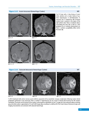
Figure 2.4.3 Acute Intracranial Hemorrhage (Canine) MR
13y FS Corgi with a 1‐day history of tonic/
clonic seizures and a 2‐year history of sys-
temic hypertension. A well‐delineated, T1
isointense (a), T2 hypointense (b) intraaxial
cerebral mass is present in the region of the
right piriform lobe. There is moderate edema
surrounding the mass (b) as well as a thin
peripheral rim of contrast enhancement (c).
There is uniform susceptibility effect within
the lesion (d).
(a) T1, TP (b) T2, TP
(c) T1+C, TP (d) T2*, TP
Figure 2.4.4 Subacute Intracranial Hemorrhage (Canine) MR
(a) T1, TP (b) T2, TP (c) FL, TP
7y MC Greyhound with central nervous system deficits associated with an anesthetic recovery complication following routine dental
prophylaxis performed 5 days previously. There is marked T1 hyperintensity (a) and mixed T2 intensity (b) involving the left cerebral
hemisphere. The lesion involves primarily the cerebral cortex based on distribution on the T1 image, but more extensive edema involving
gray and white matter is appreciated on T2 and FLAIR images (b,c), resulting in a midline shift. The 5‐day history of intracranial signs and
imaging characteristics are consistent with subacute hemorrhage.
177