
Page 186 - Atlas of Small Animal CT and MRI
P. 186
176 Atlas of Small Animal CT and MRI
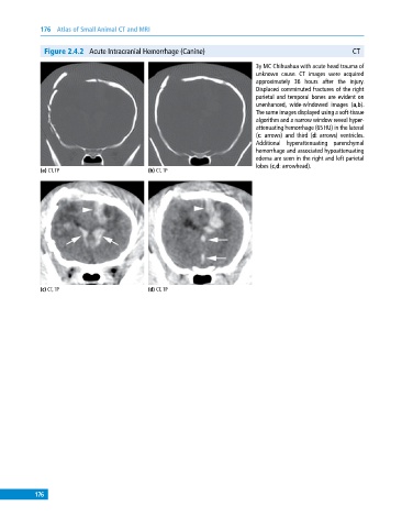
Figure 2.4.2 Acute Intracranial Hemorrhage (Canine) CT
3y MC Chihuahua with acute head trauma of
unknown cause. CT images were acquired
approximately 36 hours after the injury.
Displaced comminuted fractures of the right
parietal and temporal bones are evident on
unenhanced, wide‐windowed images (a,b).
The same images displayed using a soft‐tissue
algorithm and a narrow window reveal hyper-
attenuating hemorrhage (65 HU) in the lateral
(c: arrows) and third (d: arrows) ventricles.
Additional hyperattenuating parenchymal
hemorrhage and associated hypoattenuating
edema are seen in the right and left parietal
lobes (c,d: arrowhead).
(a) CT, TP (b) CT, TP
(c) CT, TP (d) CT, TP
176