
Page 181 - Atlas of Small Animal CT and MRI
P. 181
Developmental Disorders 171
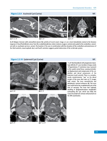
Figure 2.3.9 Arachnoid Cyst (Canine) MR
(a) T1+C, SP (b) T2, SP (c) T2, TP
4y M Belgian Tervuren with unclassified seizure‐like activity of recent onset. Image c is at a level immediately rostral to the foramen
magnum. A focal fluid dilation arises from the cerebellomedullary cistern (cisterna magna), ventral and caudal to the cerebellum, consist-
ent with an arachnoid cyst (a–c: arrow). The location of the cyst, in combination with the elevation of the cerebellum and prominence of
the third ventricle, mesencephalic duct, and fourth ventricle, suggests partial obstruction of the ventricular system.
Figure 2.3.10 Epidermoid Cyst (Canine) MR
11y M Newfoundland with progressive neuro-
logic deficits of 1‐year duration. A large, ovoid,
T2 hyperintense, T1 isointense mass is present
in the caudal cranial fossa, causing rostrodor-
sal displacement and compression of the cer-
ebellum and dorsal compression of the
brainstem (a–d: asterisk). There is a complex,
sessile mixed‐intensity “cap” on the dorsal
margin of the mass, best seen on T2 images
(a,b: arrow). The mass nonuniformly and
peripherally contrast enhances (d). The mass
was confirmed to be an epidermoid cyst at the
time of necropsy. The mass had ruptured,
resulting in lipogranulomatous encephalitis
(a) T2, SP (b) T2, TP surrounding the cyst, which was likely the
cause for peripheral contrast enhancement on
the MR examination.
(c) T1, TP (d) T1+C, TP
171