Page 177 - Atlas of Small Animal CT and MRI
P. 177

Developmental Disorders  167


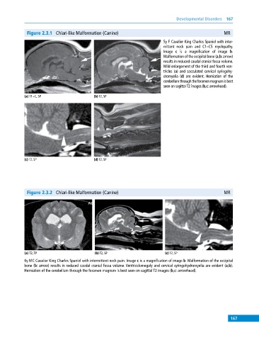
              Figure 2.3.1  Chiari‐like Malformation (Canine)                                            MR
                                                                             5y F Cavalier King Charles Spaniel with inter-
                                                                             mittent neck pain and C1–C5 myelopathy.
                                                                             Image  c is a magnification of image  b.
                                                                             Malformation of the occipital bone (a,b: arrow)
                                                                             results in reduced caudal cranial fossa volume.
                                                                             Mild enlargement of the third and fourth ven-
                                                                             tricles (a)  and sacculated cervical syringohy-
                                                                             dromyelia (d) are evident. Herniation of the
                                                                             cerebellum through the foramen magnum is best
                                                                             seen on sagittal T2 images (b,c: arrowhead).

            (a) T1+C, SP                     (b) T2, SP


            (c) T2, SP                       (d) T2, SP


              Figure 2.3.2  Chiari‐like Malformation (Canine)                                            MR


            (a) T2, TP                       (b) T2, SP                       (c) T2, SP
            6y MC Cavalier King Charles Spaniel with intermittent neck pain. Image c is a magnification of image b. Malformation of the occipital
            bone (b: arrow) results in reduced caudal cranial fossa volume. Ventriculomegaly and cervical syringohydromyelia are evident (a,b).
            Herniation of the cerebellum through the foramen magnum is best seen on sagittal T2 images (b,c: arrowhead).


                                                                                                             167
   172   173   174   175   176   177   178   179   180   181   182