
Page 178 - Atlas of Small Animal CT and MRI
P. 178
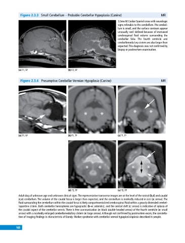
Figure 2.3.3  Small Cerebellum – Probable Cerebellar Hypoplasia (Canine)                    MR

                                                                              3.5mo M Cocker Spaniel cross with neurologic
                                                                              signs referable to the cerebellum. The cerebel-
                                                                              lum is small, and the surface contours appear
                                                                              unusually well defined because of increased
                                                                              cerebrospinal  fluid  volume  surrounding  the
                                                                              cerebellar folia.  The fourth ventricle and
                                                                                cerebellomedullary cistern are also larger than
                                                                              expected. This diagnosis was not confirmed by
                                                                              biopsy or postmortem examination.


             (a) T1, SP                       (b) T2, SP


              Figure 2.3.4  Presumptive Cerebellar Vermian Hypoplasia (Canine)                            MR


             (a) T1, SP                       (b) T1, TP                      (c) T1, TP


                                              (d) T2, TP                      (e) T2, TP
             Adult dog of unknown age and unknown clinical signs. The representative transverse images are at the level of the rostral (b,d) and caudal
             (c,e) cerebellum. The volume of the caudal fossa is larger than expected, and the cerebellum is markedly reduced in size (a: arrow). The
             fluid surrounding the cerebellum within the caudal fossa is likely compartmentalized cerebrospinal fluid within a grossly distended cerebel-
             lopontine cistern. Both cerebellar hemispheres are hypoplastic (b–e: asterisks), and the central cleft (c: arrow) is indicative of aplasia of
             the caudal aspect of the cerebellar vermis. There is free communication (e: black double‐headed arrow) of the fourth ventricle (e: small
             arrow) with a markedly enlarged cerebellomedullary cistern (e: large arrow). Although not confirmed by postmortem exam, the constella-
             tion of imaging findings is characteristic of Dandy–Walker syndrome with cerebellar vermal hypoplasia/aplasia described in people.


           168