Page 180 - Atlas of Small Animal CT and MRI
P. 180

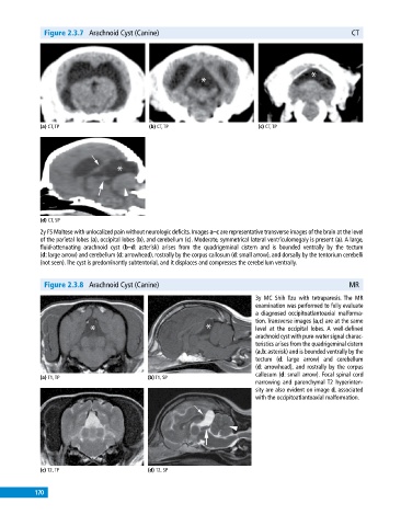
Figure 2.3.7  Arachnoid Cyst (Canine)                                                        CT


             (a) CT, TP                       (b) CT, TP                       (c) CT, TP


             (d) CT, SP
             2y FS Maltese with unlocalized pain without neurologic deficits. Images a–c are representative transverse images of the brain at the level
             of the parietal lobes (a), occipital lobes (b), and cerebellum (c). Moderate, symmetrical lateral ventriculomegaly is present (a). A large,
             fluid‐attenuating arachnoid cyst (b–d: asterisk) arises from the quadrigeminal cistern and is bounded ventrally by the tectum
             (d: large arrow) and cerebellum (d: arrowhead), rostrally by the corpus callosum (d: small arrow), and dorsally by the tentorium cerebelli
             (not seen). The cyst is predominantly subtentorial, and it displaces and compresses the cerebellum ventrally.


              Figure 2.3.8  Arachnoid Cyst (Canine)                                                       MR
                                                                              3y MC Shih Tzu with tetraparesis. The MR
                                                                              examination was performed to fully evaluate
                                                                              a diagnosed occipitoatlantoaxial malforma-
                                                                              tion. Transverse images (a,c) are at the same
                                                                              level at  the  occipital  lobes.  A  well‐defined
                                                                              arachnoid cyst with pure‐water signal charac-
                                                                              teristics arises from the quadrigeminal cistern
                                                                              (a,b: asterisk) and is bounded ventrally by the
                                                                              tectum  (d: large  arrow) and cerebellum
                                                                              (d:   arrowhead), and rostrally by the corpus
                                                                              callosum (d: small arrow). Focal spinal cord
             (a) T1, TP                       (b) T1, SP
                                                                              narrowing  and  parenchymal T2  hyperinten-
                                                                              sity are also evident on image d, associated
                                                                              with the occipitoatlantoaxial malformation.


             (c) T2, TP                       (d) T2, SP


           170
   175   176   177   178   179   180   181   182   183   184   185